Article
Hindlimb Lameness in Dogs - Differential Diagnosis & Field Management
Hindlimb lameness is a frequent presenting complaint in Indian veterinary clinics, especially among large-breed dogs. Accurate diagnosis and effective field management of this issue require a structured approach to differentiating common orthopedic conditions such as cranial cruciate ligament rupture (CCLR), hip dysplasia, patellar luxation, and musculoskeletal trauma.
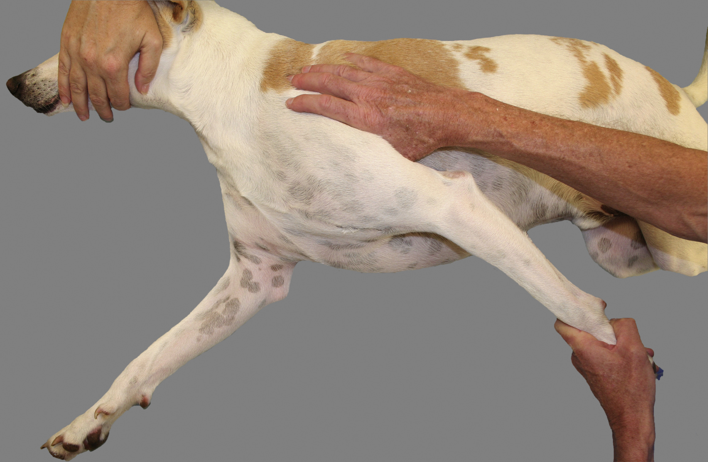
Cranial cruciate ligament rupture is the leading cause of hindlimb lameness in adult dogs. It is commonly seen in Labradors, German Shepherds, and Indian Indies. Dogs typically present with acute, non-weight-bearing lameness and stifle joint effusion. Key diagnostic tests include the cranial drawer and tibial thrust tests. Radiographs may reveal joint effusion and osteophytes in chronic cases (Duerr et al., 2016).
Hip dysplasia, often bilateral and genetically predisposed, affects the coxofemoral joint and presents as bunny-hopping gait, difficulty rising, and muscle wasting. Diagnosis requires sedation and proper radiographic positioning (extended VD view). The Ortolani sign can be helpful in detecting hip laxity in young dogs.
Patellar luxation, particularly medial luxation, is more common in small breeds but can be seen in medium-breed Indian dogs. Clinical signs include intermittent skipping lameness and reluctance to jump. Diagnosis is manual palpation often reveals spontaneous luxation
during flexion (Innes et al., 2000).
In field setups with limited imaging access, a tiered management approach is essential(Di Dona et al., 2018):
- Start with cage rest (5–7 days), cold compress, and NSAIDs (e.g., carprofen 4.4 mg/kg SID).
- Monitor response and re-evaluate lameness at day 7–10.
- Persistent or non-improving cases should be referred for orthopedic consultation and imaging.
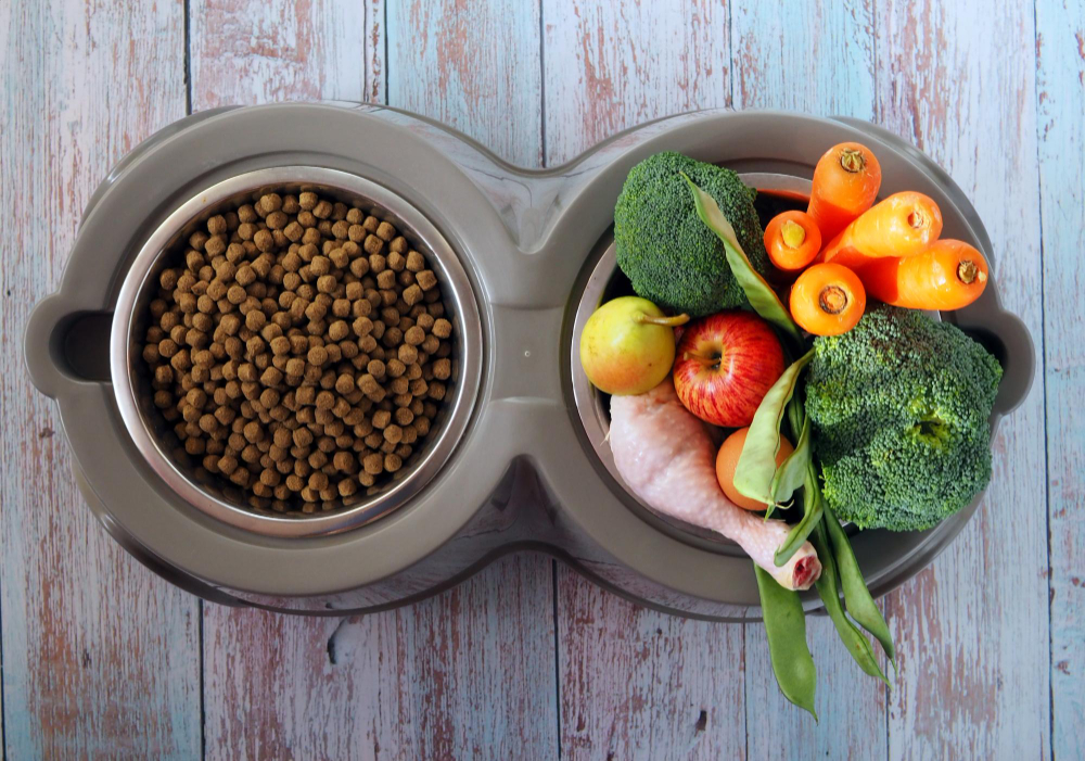
Rehabilitation exercises such as assisted walking, weight shifts, and underwater treadmill therapy improve recovery post-diagnosis or surgery. Owner education on exercise restriction, dietary control, and joint supplementation (e.g., omega-3s, glucosamine) is crucial to avoid recurrence.
References:
- Duerr FM et al. (2016). https://pubmed.ncbi.nlm.nih.gov/25328024/
- Innes JF & Bacon D. (2000). https://pubmed.ncbi.nlm.nih.gov/11058021
- Di Dona, F., Della Valle, G., & Fatone, G. (2018). https://doi.org/10.2147/vmrr.s142545
Related Contents
Upcoming Event
test ev
test
Article
test
test
Article
dfgdfhfhd
fhfhfd
Video
test
Good leadership is necessary for the success of every company. The health care industry, including t...
Video
xcncxnxcn
cbbxzbxzb
Article
test
Word Documents Template Main heading: Use the Heading 1 style for primary...
Article
test test footnote
[2]Test [3]footnote[1]Reference(s):1. Test 22. Test 33. Test test 3
Article
Test New Editor
1Precision Animal Nutrition (PAN) as a novel Tool in the Feeding of Animals in India The future...